
From Wikipedia, the free encyclopedia
This article is about infection of skin and its underlying connective tissue. For the dimpled appearance of skin, see cellulite.
| Cellulitis | |
|---|---|
| Classification and external resources | |

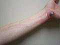
Infected skin with cellulitis
|
|
| ICD-10 | L03 |
| ICD-9 | 682.9 |
| DiseasesDB | 29806 |
| MedlinePlus | 000855 |
| eMedicine | med/310 emerg/88 derm/464 |
| MeSH | D002481 |
Erysipelas is the term used for a more superficial infection of the dermis and upper subcutaneous layer that presents clinically with a well-defined edge. Erysipelas and cellulitis often coexist, so it is often difficult to make a distinction between the two.
In Ludwig's angina, an acute and potentially life threatening condition, cellulitis occurs within the submandibular (lower jaw) space.[1]
Cellulitis is unrelated (except etymologically) to cellulite, a cosmetic condition featuring dimpling of the skin.
Contents
Signs and symptoms
The typical symptoms of cellulitis is an area which is red, hot, and painful. The photos shown here of cellulitis are of mild cases, and are not representative of earlier stages of the condition.-
 Cellulitis following an abrasion. Note the red streaking up the arm from involvement of the lymphatic system.
-
 Infected left shin in comparison to shin with no sign of symptoms
-
 Cellulitis of the leg with foot involvement.


Causes
Cellulitis is caused by a type of bacteria entering the skin, usually by way of a cut, abrasion, or break in the skin. This break does not need to be visible. Group A Streptococcus and Staphylococcus are the most common of these bacteria, which are part of the normal flora of the skin, but normally cause no actual infection while on the skin's outer surface.Dental infections account for approximately 80% of cases of Ludwig's angina, or cellulitis of the submandibular space. Mixed infections, due to both aerobes and anaerobes, are commonly associated with the cellulitis of Ludwig's angina. Typically this includes alpha-hemolytic streptococci, staphylococci and bacteroides groups.[1]
Predisposing conditions for cellulitis include insect or spider bite, blistering, animal bite, tattoos, pruritic (itchy) skin rash, recent surgery, athlete's foot, dry skin, eczema, injecting drugs (especially subcutaneous or intramuscular injection or where an attempted intravenous injection "misses" or blows the vein), pregnancy, diabetes and obesity, which can affect circulation, as well as burns and boils, though there is debate as to whether minor foot lesions contribute.
Occurrences of cellulitis may also be associated with the rare condition hidradenitis suppurativa.
The appearance of the skin will assist a doctor in determining a diagnosis. A doctor may also suggest blood tests, a wound culture or other tests to help rule out a blood clot deep in the veins of the legs. Cellulitis in the lower leg is characterized by signs and symptoms similar to those of a deep vein thrombosis, such as warmth, pain and swelling (inflammation).
This reddened skin or rash may signal a deeper, more serious infection of the inner layers of skin. Once below the skin, the bacteria can spread rapidly, entering the lymph nodes and the bloodstream and spreading throughout the body. This can result in influenza-like symptoms with a high temperature and sweating or feeling very cold with shaking, as the sufferer cannot get warm.
In rare cases, the infection can spread to the deep layer of tissue called the fascial lining. Necrotizing fasciitis, also called by the media "flesh-eating bacteria", is an example of a deep-layer infection. It is a medical emergency.
Risk factors
The elderly and those with immunodeficiency (a weakened immune system) are especially vulnerable to contracting cellulitis. Diabetics are more susceptible to cellulitis than the general population because of impairment of the immune system; they are especially prone to cellulitis in the feet, because the disease causes impairment of blood circulation in the legs, leading to diabetic foot/foot ulcers. Poor control of blood glucose levels allows bacteria to grow more rapidly in the affected tissue, and facilitates rapid progression if the infection enters the bloodstream. Neural degeneration in diabetes means these ulcers may not be painful and thus often become infected. Those who have suffered poliomyelitis are also prone because of circulatory problems, especially in the legs.Immunosuppressive drugs, and other illnesses or infections that weaken the immune system, are also factors that make infection more likely. Chickenpox and shingles often result in blisters that break open, providing a gap in the skin through which bacteria can enter. Lymphedema, which causes swelling on the arms and/or legs, can also put an individual at risk.
Diseases that affect blood circulation in the legs and feet, such as chronic venous insufficiency and varicose veins, are also risk factors for cellulitis.
Cellulitis is also extremely prevalent among dense populations sharing hygiene facilities and common living quarters, such as military installations, college dormitories, nursing homes, oil platforms and homeless shelters. It is advised if a cabin is shared with a sufferer, urgent medical treatment should be given.
Diagnosis
Cellulitis is most often a clinical diagnosis, and local cultures do not always identify the causative organism. Blood cultures usually are positive only if the patient develops generalized sepsis. Conditions that may resemble cellulitis include deep vein thrombosis, which can be diagnosed with a compression leg ultrasound, and stasis dermatitis, which is inflammation of the skin from poor blood flow. Associated musculoskeletal findings are sometimes reported. When it occurs with acne conglobata, hidradenitis suppurativa, and pilonidal cysts, the syndrome is referred to as the follicular occlusion triad or tetrad.[2]Lyme disease can be misdiagnosed as staphylococcal- or streptococcal-induced cellulitis. Because the characteristic bullseye rash does not always appear in patients infected with Lyme disease, the similar set of symptoms may be misdiagnosed as cellulitis. Standard treatments for cellulitis are not sufficient for curing Lyme disease. The only way to rule out Lyme disease is with a blood test, which is recommended during warm months in areas where the disease is endemic.[3]
No comments:
Post a Comment